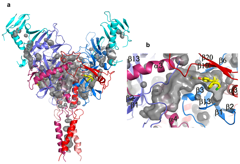
Extended Data Figure 4. Pockets and tunnels in EBOV GP trimer.
a, The several small pockets and three large tunnels in the GP trimer shown as grey surfaces. Protein backbones are drawn as ribbons and coloured as in Fig. 2 of the main text. A toremifene is bound at the entrance of each large tunnel and shown as yellow sticks. b, Close up view of a tunnel. Each tunnel is bordered by secondary structure elements from two neighbouring monomers.

