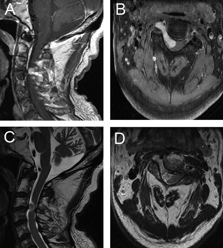
Fig. 1.

Preoperative imaging in a patient with cervical myelopathy of nondegenerative pathology. An 80-year-old man presented with 6-month history of bilateral upper extremity weakness and numbness. (A, B) T1-weighted magnetic resonance imaging with contrast revealed an enhancing lesion extending from the neural foramen into the spinal canal. (C, D) T2-weighted magnetic resonance imaging was notable for significant cord compression and the absence of hyperintensity in the spinal cord. The patient was taken to the operating room for resection of this lesion, and pathology confirmed the diagnosis of schwannoma.
