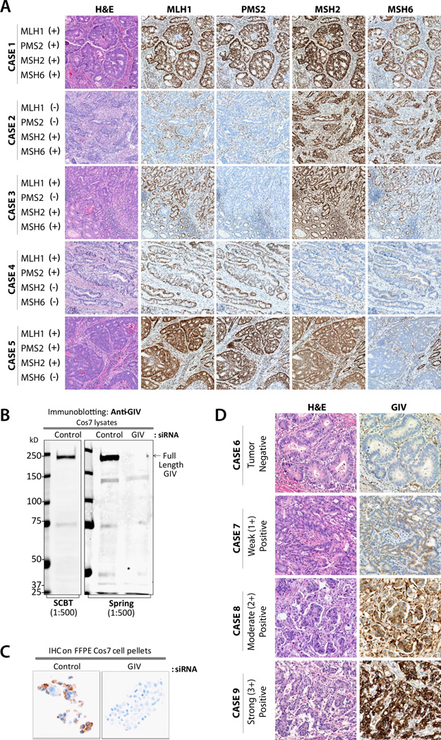
Figure 1. Immunohistochemical staining of primary colon tumors for mismatch repair (MMR) proteins and GIV.
(A) A proficient MMR case (Case 1) shows intact nuclear staining for all four MMR proteins in tumor and stromal tissue. Deficient MMR cases (Cases 2–5) represent loss of MLH1, PMS2, MSH2, and/or MSH6 proteins, respectively, in tumor cells, while the intact nuclear staining is preserved in stromal cells or normal colonic epithelium. (B) Equal aliquots (~ 75 ug) of whole cell lysates of control or GIV-depleted Cos7 cells were analyzed for GIV expression by immunoblotting with anti-GIV antibodies as indicated. (C) Pellets of control and GIV-depleted Cos7 cells were fixed, embedded in paraffin and analyzed for GIV expression by IHC with anti-GIV antibody. (D) A representative case (Case 6) negative for GIV expression. Cases 7–9 demonstrate weak, moderate, and strong GIV expression, respectively, in tumor cells.

