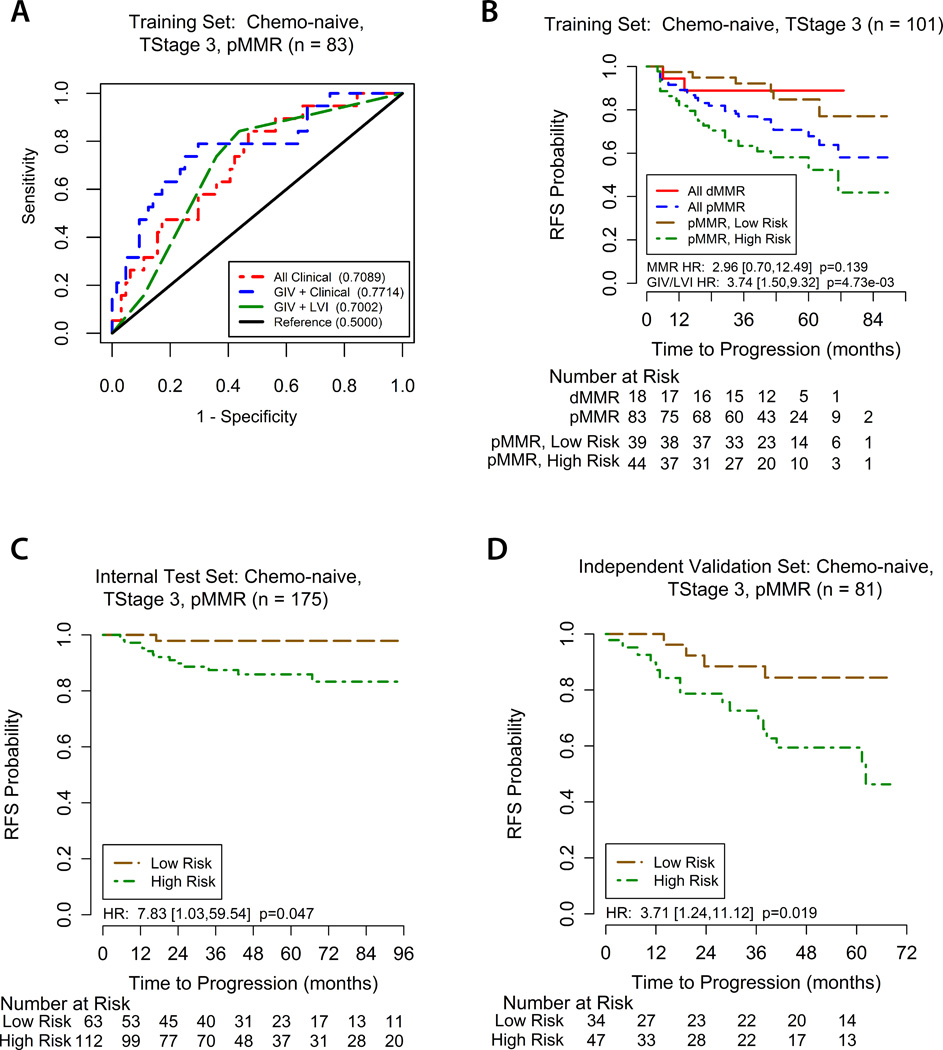
Figure 2. Assessment of GIV/LVI risk model.
(A) ROC curves comparing the prognostic accuracy of the GIV/LVI risk classifier (high vs low risk) with clinical model alone, or GIV and clinical model combined. Area under the curve (AUC) for recurrence at 3 years (shown in brackets) shows the utility of including GIV analysis in recurrence risk assessment. (B) Kaplan-Meier recurrence-free survival (RFS) based on MMR status and the GIV/LVI risk classifier for T3, surgery-alone patients in the training set. (C, D) Kaplan-Meier RFS based on the GIV/LVI risk classifier for T3, pMMR, surgery-alone patients in the internal testing (C) and independent validation sets (D). ROC = receiver operator characteristics. LVI = lymphovascular invasion. dMMR/pMMR = deficient/proficient mismatch repair.

