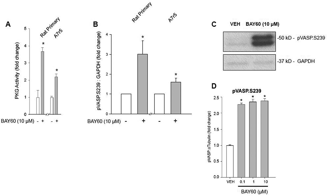
Figure 5. BAY60 increases PKG activity and VASP S239 phosphorylation.
(A) Treatment with BAY60 (10 µM, 60 min) stimulated significant increases in PKG activity in rat primary and rat A7r5 ASM cells compared to VEH controls assessed through a PKG activity ELISA. (B–C) Densitometric quantification of rat primary, and rat A7r5 cell homogenates along with a representative ECL Western blot for VASP (at the preferred PKG site, Ser239 (pVASP.S239)) and GAPDH. Data reveal significant increases in pVASP.S239 compared to respective VEH controls for each cell type. (D) Densitometric quantification from an In-Cell Western blot using intact, adherent rat primary ASM cells showing BAY60 significantly increases pVASP.S239 levels (normalized to α-tubulin) compared to VEH controls. n ≥ 3 independent experiments for PKG activity, ECL and ICW Western blots. * p < 0.05 vs. VEH controls.

