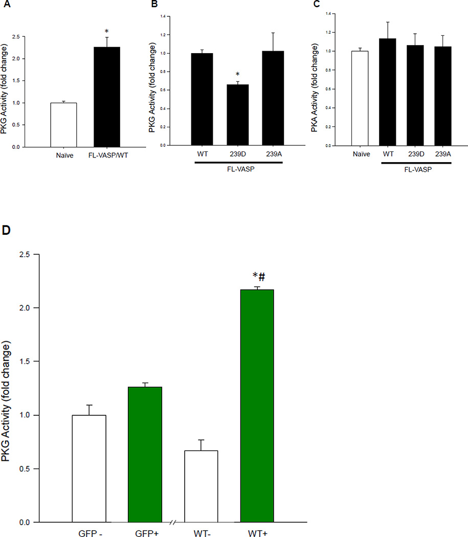
Figure 8. FL-VASP mutants differentially regulate PKG activity.
(A) FL-VASP/ WT significantly increases PKG activity compared to naïve controls 48 hours post-transfection using a PKG activity ELISA. (B) At this same time point, phospho-mimetic FL-VASP/239D prevented FL-VASP-mediated enhancement of PKG activity compared to FL-VASP/WT and phospho-resistant FL-VASP/239A overexpression. These changes were observed in the absence of any significant increases in PKA activity (C). (D) In order to confirm these findings, lysates from the previously described FACS sort (Figure 6E) were probed for PKG activity and results showed FL-VASP/WT or “WT+” cells significantly increased PKG activity compared to all control groups. (A) n=2 (B–C) n=3 (D) n=1 independent experiment(s) performed in quadruplicate. * p < 0.05 vs. naïve (A,C) or WT (B) or GFP+ (D); (D) # p < 0.05 vs. WT−.

