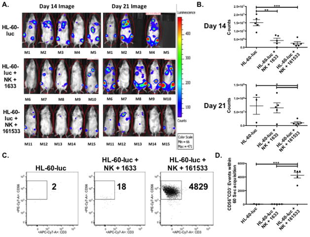
Figure 6. 161533 TriKE limits HL-60 tumor growth in vivo better than 1633 BiKE through enhanced maintenance of NK cells.
An in vivo tumor model was established by conditioning NSG mice (275 cGy) and then injecting HL-60-luc cells intravenously (7.5 × 10⁵ cells/mouse). Three days later 1×10⁶ normal human donor NK cells (calculated from a magnetically depleted CD3/CD19 product) activated overnight with IL-15 were infused. 1633 BiKE and 161533 TriKE (50 ug) was administered MTWThF through the next two weeks of the study (10 doses total), and a control group only received HL-60-luc cells. A) Individual mouse photoluminscence after 2 minute exposures on day 14 (left) and day 21 (right) after NK infusion are shown (n=5 per treatment group,). B) Quantification of luminescence in mice from the three treatment groups at day 14 (top) and day 21 (bottom). Each dot represents a different mouse and bars denote mean ± SEM (n=5 per treatment group, representative of two separate experiments). C) Blood was collected on day 20 from the mice in each of the experiential treatment groups. Circulating CD45+CD56+CD3− human NK cells were quantified by flow cytometry. Events were collected over 60 seconds and the number of human NK cell events was calculated. Representative dot plots are shown denoting the number of NK (CD56+CD3−) cell events within the CD45+ gate. D) Aggregate data demonstrating the number of human NK cell events in each treatment group at day 20. Individual dots represent different mice and bars denote mean ± SEM (n=3 for HL-60-luc group [two mice died], n=5 for the 1633 BiKE and 161533 TriKE groups).

