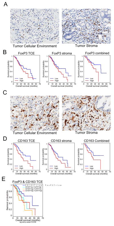
Figure 1. Immunosuppressive cells are associated with poor outcome in PDA.
(A) Representative images of High FoxP3 staining in the tumor cellular environment (TCE, >0%) and stroma (>9.5%) of patient sections. (B) Kaplan-Meier plots indicating survival probability in human patients with High or Low accumulation of FoxP3+ cells in the tumor cellular environment and/or stroma. See supplementary Table S1,S2. (C) Representative images of High CD163 staining in the tumor cellular environment (>30%) and stroma (>33%) of patient sections. (D) Kaplan-Meier plots indicating survival probability in human patients with High or Low accumulation of CD163+ cells in the tumor cellular environment and/or stroma. See supplementary Table S3,S4. (E) Kaplan-Meier plots indicating survival probability in human patients with High or Low accumulation of CD163+ and FoxP3+ cells in the tumor stroma. See supplementary Table S5.

