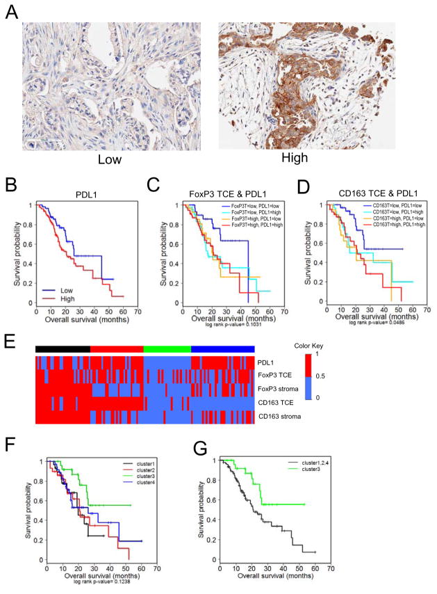
Figure 3. Immune evasion and suppression signature correlates with poor prognosis in PDA.
(A) Representative images of Low (<10%) and High (≥10%) PD-L1 staining of patient PDA tumor sections. (B) Kaplan-Meier plots indicating survival probability in human patients with High or Low accumulation of PD-L1+ cells. See supplementary Table S6, S7. (C) Kaplan-Meier plots indicating survival probability in human patients with High or Low accumulation of FoxP3+ and PD-L1+ cells in the tumor cellular environment (TCE). See supplementary Table S8. (D) Kaplan-Meier plots indicating survival probability in human patients with High or Low accumulation of CD163+ and PD-L1+ cells in the tumor cellular environment. See supplementary Table S9. (E) Heat maps of unsupervised RF clustering of immune marker expression. Clusters were defined using the partitioning around medoids method. (F) Kaplan-Meier plots indicating survival probability in human patients based on the previously defined immune marker expression cluster. See supplementary Table S10. (G) Kaplan-Meier plots indicating survival probability in human patients from the cluster of lowest complex expression (cluster 3) with those from clusters with high expression of at least one marker (clusters 1,2,4). See supplementary Table S11.

