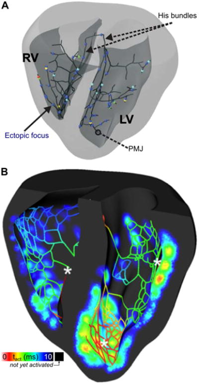
Fig. 2.

Premature ventricular contractions (PVCs) originating in the Purkinje system (PS). (A) 3D map showing locations of initial excitation that triggered PVCs in a model of the rabbit PS and ventricles. All cells in the model were prone to delayed afterdepolarization (DAD)-induced excitation due to spontaneous calcium release, but such activity occurred exclusively in the PS due to lower source-sink mismatch. PMJ = Purkinje-myocardial junction. (Reprinted with permission from Ref (Campos et al., 2015)) (B) 3D map showing activation times in a cutaway view of the rabbit ventricles and PS during a post-pause propagating response caused by DAD. Asterisks show locations where DADs occurred in the PS, leading to propagating excitation that eventually caused a PVC. (Reprinted with permission from (Zamiri et al., 2014))
