
Fig. 4.
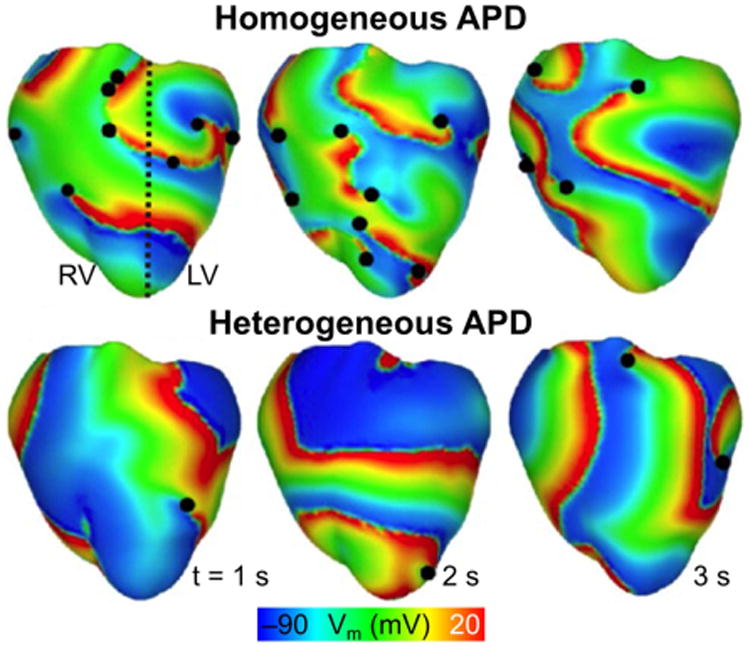
Role of LV/RV APD heterogeneity on VF dynamics. Transmembrane potential distributions during VF for model with heterogeneous APD (i.e. left, LV, and right, RV, ventricles have different APDs) and for model with homogenous APD. The dashed black line denotes the border between regions characterized by a different APD. Epicardial phase singularities are marked with solid black circles. (Modified and reprinted with permission from Ref (Arevalo et al., 2007))