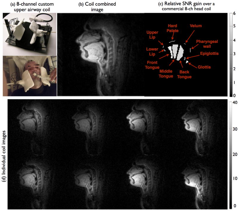
Figure 2.
The custom eight channel upper-airway coil and its placement on a subject (a). As shown in (d), the individual coil images from all the channels depict high signal on all relevant upper-airway regions. (b) depicts the coil combined image that demonstrate high SNR on all the upper-ariway regions. (c) depicts the relative SNR gain map over a commercial eight-channel head coil, where SNR gains between 2 to 6 fold are observed in all the upper-airway regions of interest.

