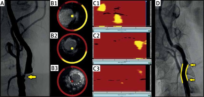
Figure 1.
The angiogram of the left carotid artery revealed a 90% stenosis for which stenting was indicated (A). The spectroscopic image obtained prior to the intervention revealed two distinct lipid cores located proximally and distally to the critical 90% stenosis of the left internal carotid artery (C1). The corresponding IVUS image is provided (B1). The lipid cores disappeared after stent deployment (C2) and subsequent post-dilation of the lesion (panel C3). The corresponding IVUS images can be seen in panels B2 and B3. Final carotid angiography confirmed a good result (D). The stented area is marked in yellow

