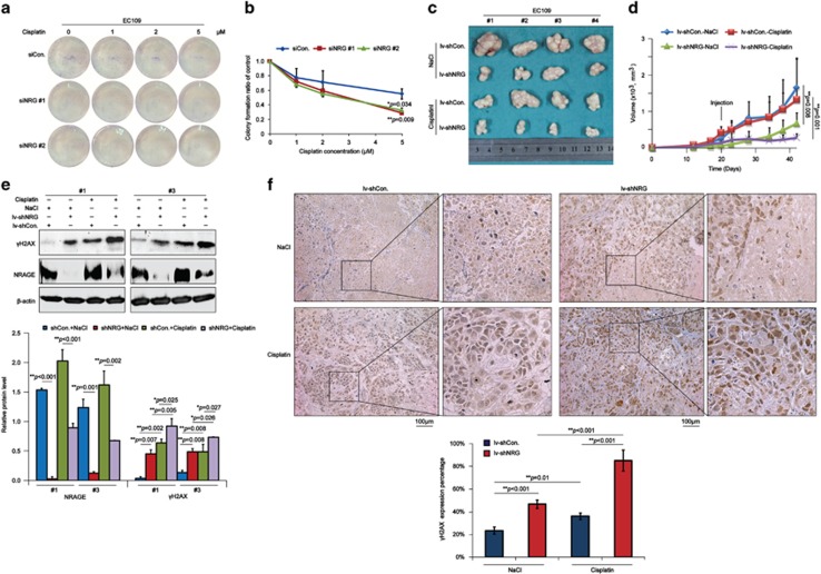
Figure 2.
NRAGE knockdown sensitizes EC cells to cisplatin both in vitro and in vivo. (a) EC109 cells transfected with siCon. or siNRG (#1, #2) were treated with the indicated concentrations of cisplatin for 2 h and left to form the colonies for 14 days. (b) The number of colonies with >50 cells in panel (a) was manually counted, followed by normalization to EC109 cells transfected with siCon. or siNRG (#1, #2) without cisplatin treatment, respectively. Data were represented as means±S.D. (*P=0.034 for siNRG #1, **P=0.009 for siNRG #2). (c) EC109/lv-shCon. and EC109/lv-shNRG cells were subcutaneously transplanted into male nude mice (N=4), and the mice were subsequently intraperitoneally injected with 0.9% NaCl or 10 mg/kg cisplatin when the bearing tumors were about 200 mm3. Finally, tumors were dissected and pictured. (d) Tumor volumes in panel (c) were measured and statistically analyzed (**P=0.008 for the NaCl group, **P=0.001 for the cisplatin group). Error bar represents S.D. (e) Tumor tissues from #1 and #3 in panel (c) were lysed and subjected to western blotting assays with the indicated antibodies. The gray values of western bands from three independent experiments were used for the ANOVA statistical analysis. (f) Tumors in panel (c) were detected of γH2AX using IHC assays (scale bar is 100 μm). The expression percentage of γH2AX was blindly evaluated by a professional doctor and finally used for the ANOVA statistical analysis

