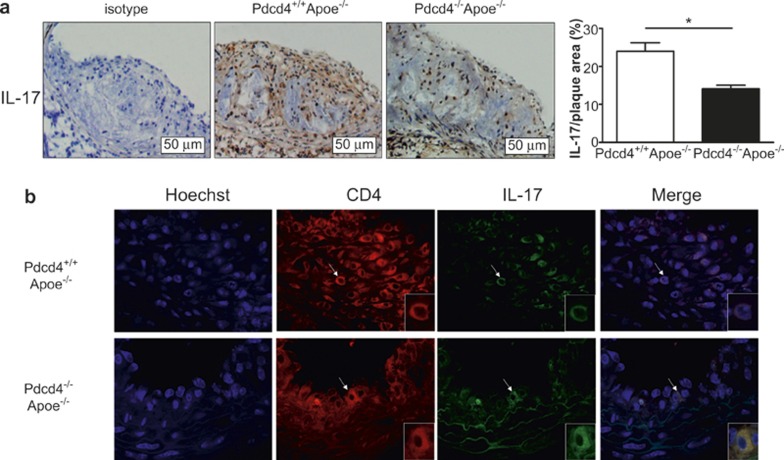
Figure 4.
A low level of IL-17 was expressed in the atherosclerotic lesions of the Pdcd4−/−Apoe−/− mice. The Pdcd4+/+Apoe−/− and Pdcd4−/−Apoe−/− mice were fed a high fat diet from the age of 8 weeks to 16 weeks. The aortic roots with their atherosclerotic plaques were isolated to prepare serial frozen sections. (a) IHC staining of serial frozen sections using an anti-IL17 Ab revealed IL-17 expression primarily in the atherosclerotic plaques (original magnification ×200). The data are shown as the means ± SEM. *P < 0.05. (b) The cell nuclei were stained blue with Hoechst, and IL-17 and CD4 proteins were co-localized in the same section by dual IF staining. CD4 (in red) was stained with rat anti-mouse antibody and TRITC-conjugated anti-rat second antibody, and IL-17 (in green) was stained with rabbit anti-mouse antibody and FITC-conjugated anti-rabbit second antibody. The merged image shows co-expression of IL-17 and CD4 in the same cells (yellow). The cell indicated by the white arrows is shown magnified in the lower right corner. The data are derived from four Pdcd4+/+Apoe−/− mice and five Pdcd4−/−Apoe−/− mice.

