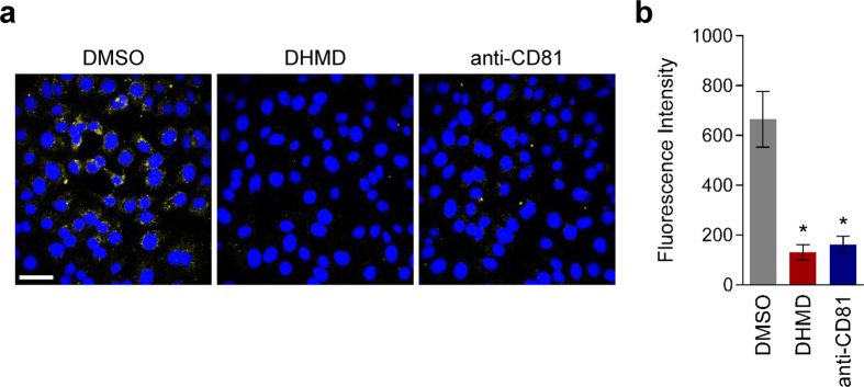
Figure 7. Confocal microscopy analysis of DHMD’s inhibitory effect against HCV adsorption.
(a) Huh-7.5 cells seeded in chamber wells were co-incubated with HCVcc (MOI = 0.5) and DHMD (50 μM) or the controls at 4 °C for 3 h, then washed with PBS before shifting to 37 °C for 3 h. At the end of the incubation, cells were washed and fixed for immunostaining using anti-HCV core antibody and visualized with a confocal microscope. Nuclei were stained with the mounting medium used, which contained DAPI. DMSO = 0.5%; anti-CD81 = 10 μg/ml; magnification = 60×; scale bar = 50 μm. Data shown are means ± SEM (*P < 0.05) or representative micrographs from 3 independent experiments.

