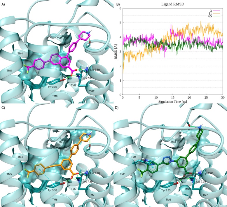
Figure 2.
(A) Docking pose of reference compound 3 (magenta-colored carbons) at hP2Y14R. (B) RMSD plots for the considered compounds during 30 ns of membrane MD simulations. (C) Docking pose of the alkynyl derivative 11 (orange-colored carbons) at hP2Y14R. (D) Docking pose of the triazolyl derivative 65 (green-colored carbons) at the hP2Y14R. Side chains of residues important for ligand recognition are reported as sticks (dark-cyan carbon atoms). Side chains of residues establishing either van der Waals or hydrophobic contacts with the ligand are rendered as transparent surface. H-Bonds are pictured as green solid lines, whereas π–π stacking interactions as cyan dashed lines with the centroids of the aromatic rings displayed as cyan spheres. Nonpolar hydrogen atoms are omitted.

