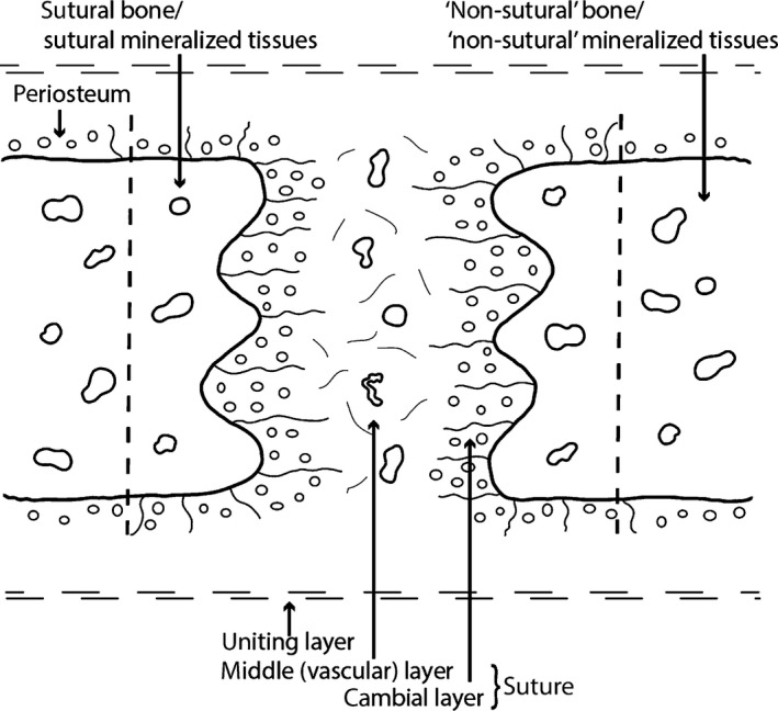
Figure 1.

Schematic representation of a mammalian suture. Sutures are composed of a cambial layer with numerous osteoblasts and a vascular middle layer (non‐osteogenic). The sutural cambial layer is continuous with the periosteum of the bones. In the present study, the mineralized tissues directly bordering the sutures are referred to as sutural bone or sutural mineralized tissues. The mineralized tissues that are more distant from the sutural borders are referred to as ‘non‐sutural’ bone (or ‘non‐sutural’ mineralized tissues). Skull bones and their sutures are united together by the uniting layers, dense regular connective tissues mostly composed of collagen fibers. This figure is a re‐interpretation of the drawings of Pritchard et al. (1956) and Persson (1973).
