
Figure 6.
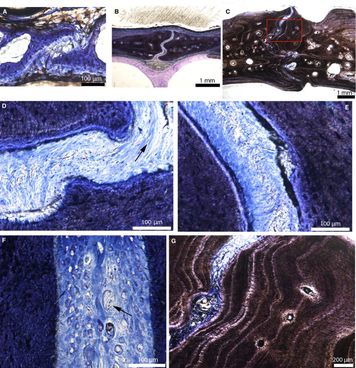
Cross‐sections of the internasal suture of American alligators stained with toluidine‐blue. (A) Internasal suture of Alligator 1. (B) Internasal suture of Alligator 3. (C) Internasal suture of Alligator 6. (D) Close‐up of the suture in (B), showing one uniform fibrous layer. Fibers can be oriented perpendicular or parallel to the sutural borders (black arrow). This suture lacks a periosteal cambial layer. (E) Close‐up of another area in the suture in (B), showing fibers that are perpendicular to the sutural borders. The suture is composed of a uniform dense fibrous connective tissue with fibroblast nuclei stained in blue. Again it lacks a periosteum. (F) Ectocranial part of the internasal suture of Alligator 4. The suture appears to be filled in with many nerves (black arrow). (G) Close‐up of the red box in (C). The sutural borders are composed of lamellae of fibrous bone, all parallel to the suture. This resembles lamellar‐zonal bone. Images (A and B) are modified from Bailleul et al. (in press).