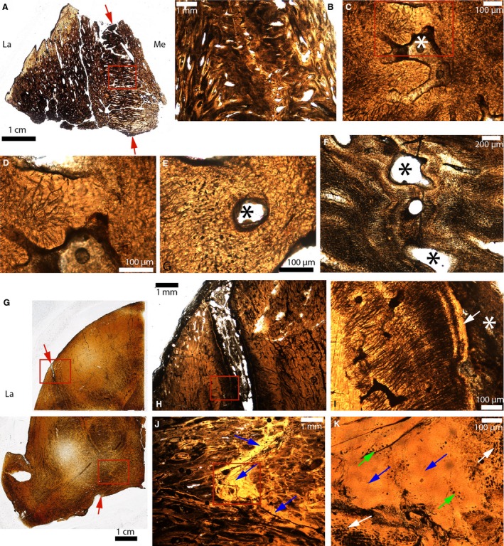
Figure 10.

Paleohistological cross‐sections in the parietal‐postorbital sutures of some pachycephalosaurids. (A) Whole section of a broken dome fragment of a Pachycephalosauridae indet (MOR 2915). The ectocranial and endocranial sides of the parietal‐postorbital suture are indicated by the red arrows. (B) Close‐up of the red box in (A). (C) Close‐up of (B) showing an obliterated suture. The fused bony fronts are fibrous and acellular. The white asterisk indicates what might be bone marrow and/or vascularized spaces. (D) Close‐up of the red box in (C). (E) Close‐up of another obliterated area of this same suture. Here, the tissue looks fibrous and cellular. The black asterisk indicates what might be bone marrow and/or vascularized spaces. (F) Another area of this same suture where the sutural fronts are extremely fibrous and parallel to the sutural borders. The black asterisk indicates what might be bone marrow and/or vascularized spaces. (G) Whole sections of half of a Pachycephalosaurus dome (VRD 13). The ectocranial and endocranial sides of the parietal‐postorbital suture are indicated by the red arrows. (H) Close‐up of the upper red box in (G). The ectocranial part of this suture is open. (I) Close‐up of the red box in (H). The sutural borders are composed of an acellular tissue (white arrow). The suture is indicated by the white asterisk. (J) Close‐up of the lower red box in (G), showing an interdigitated suture completely obliterated by a bright yellow acellular tissue (blue arrows). (K) Close‐up of the red box in (J). At higher magnification, this tissue has zones with regular and numerous osteocyte lacunae (white arrows), zones with lacunae that are less numerous and less regular (green arrows) and acellular areas (blue arrows). La, lateral; Me, medial.
