
Figure 11.
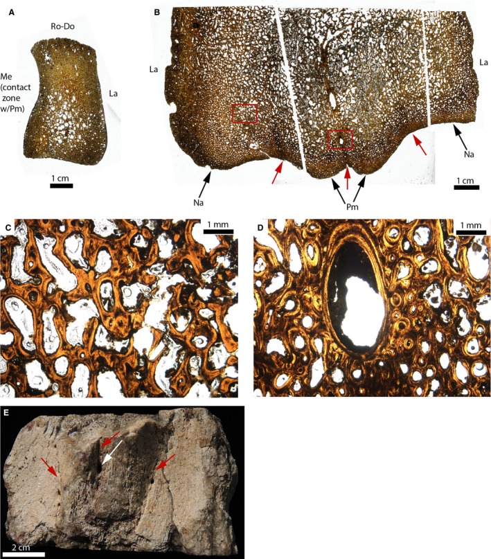
Paleohistological cross‐sections of an isolated nasal (A), and of the fused nasals and premaxillae of Triceratops (B–E). (A) Whole section of an unfused nasal (MOR 2587). (B) Whole sections of the ventral part of fused premaxillae and nasals (MOR 8661). The ventral part of the two nasal‐premaxilla and the interpremaxillary sutures are indicated by the red arrows. (C) Close‐up of the left red box in (B). It shows no trace of the suture. It is obliterated and replaced by lamellar bone trabeculae, and vascular and/or bone marrow spaces. (D) Close‐up of the right red box in (B), showing a canal present at the interpremaxillary suture. The suture is also obliterated here. (E) Photograph of the morphology of these elements in ventral view. The canal is indicated by the white arrow. The three sutures are obliterated morphologically but their general locations are indicated by the red arrows. La, lateral; Me, medial; Na, nasal; Pm, premaxilla; Ro‐Do, rostro‐dorsal.