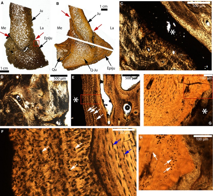
Figure 12.

Paleohistological cross‐sections of the jugal‐epijugal and the jugal‐quadratojugal sutures of Triceratops. (A) Whole section of UCMP 174838. The lateral and medial parts of the suture are indicated by the red arrows. (B) Whole sections of MOR 2570. The lateral and medial parts of the suture are indicated by the red arrows. (C) Close‐up of the suture in (A). The sutural borders on the lower part of the picture are composed of a very fibrous tissue. The suture is indicated by the white asterisk. (D) Close‐up of the red box in (A). The same fibrous tissue as that presented in (C) is also found at many external borders. It has previously been designated as ‘metaplastic bone’. (E) Close‐up of another part of this same suture, showing lamellar bone on the right and some lamellae that are parallel to the suture (white arrows). The suture is indicated by the white asterisk. (F) Close‐up of the red box in (E), showing the internal lamellar bone with regular osteocyte lacunae (blue arrows) and a tissue composed of irregular dark spaces (white arrows) near the suture. (G) Close‐up of the suture in (B). The sutural borders are composed of an acellular tissue. The white asterisk indicates the suture. (H) Close‐up of the red box in (G). The white arrows indicate cellular lacunae of various shapes and sizes. Abbreviations: same as previous figures; Epiju, epijugal; Ju, jugal; Q‐Ju, quadratojugal; Qu, quadrate.
