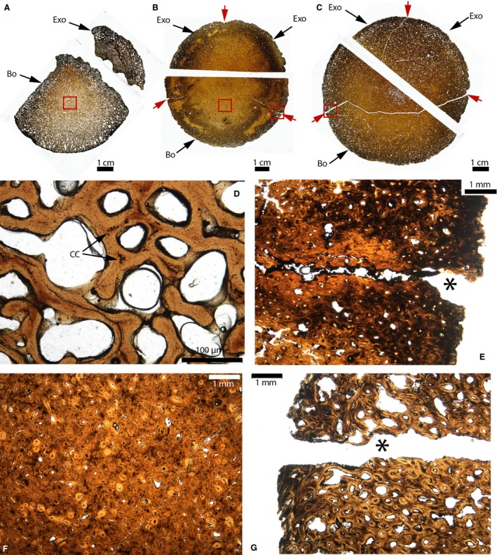
Figure 16.

Paleohistological cross‐sections of the occipital condyles of Triceratops. (A) Whole sections of the unfused occipital condyle of MOR 1110. (B) Whole sections of the fused occipital condyle of MOR 8657. (C) Whole sections of the fused occipital condyle of MOR 8658. Synchondroses are indicated by the red arrows in (B and C). (D) Close‐up of the red box in (A). Trabeculae of lamellar bone can be seen, as well as some islands of calcified cartilage, remnants of the primary cartilage model (black arrows). (E) Close‐up of the right red box in (B). The right basioccipital‐exoccipital synchondrosis is open externally (black asterisk) and does not present any calcified cartilage. It presents the histological charateristics of a suture. (F) Close‐up of the left red box in (B). The bone is a compacta with numerous secondary osteons. (G) Close‐up of the red box in (C). It presents the same histological characteristics as (E). Abbreviations: same as previous figures; CC, calcified cartilage.
