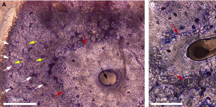
Figure 18.

Cross‐sections of a mineralized tendon from M. extensor carpi radialis of Bubo virginianus (Great Horned Owl). (A) Part of a mineralized tendon of MOR.H.2001‐10R. A vascular canal can be seen (black arrow). It does not show the typical characteristics of a primary osteon or a haversian system, and therefore was named ‘secondary reconstruction’ (SR) by Horner et al. (2015). The boundary between this SR and the rest of the tissue is ragged and undistinct (red arrows). It appears to be made of small collagen fiber bundles and fascicles tightly packed together. Further away from this SR, the tissue is composed of larger fiber fascicles (yellow arrows), separated from each other by spaces of different shapes, many of which are arc‐shaped (white arrows). These arc‐shaped spaces are the endotenons of the fiber fascicles. No regular osteocyte lacunae (nor canaliculi) can be seen anywhere in this tendon. (B) Section of another area in this same tendon showing the boundary of a SR where large fascicles are deconstructed into smaller fascicles (red arrows). This figure is modified from Horner et al. (2015).
