
Figure 1.
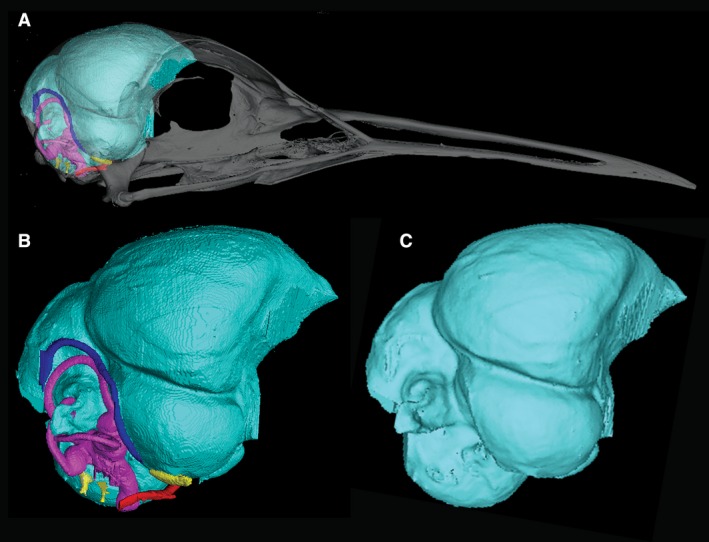
Three‐dimensional (3D) renderings of the cranial endocast of Eurypyga helias constructed from CT data. (A) Cranial endocast of E. helias with transparent skull. (B) Cranial endocast of E. helias with cranial nerves (yellow), arteries (red), veins (blue), and inner ear (pink). (C) Cranial endocast of E. helias with inner ear, nerves, and vasculature digitally removed. A smoothing algorithm (Laplacian Smooth) has been applied to the rendering.