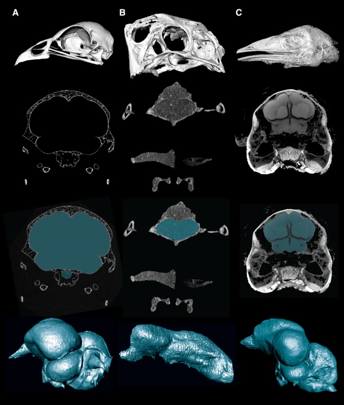
Figure 2.

CT images and cranial endocasts from different specimen types and preparations. Top row, 3D reconstruction of skull/head from CT data. Second row, representative CT slice through the cranial cavity. Third row, CT slice with the cranial cavity segmented. Fourth row, digitally prepared cranial endocast. (A) A skeletal preparation of the extant galliform bird Alectura lathami. (B) A fossil oviraptorosaur dinosaur, Citipati osmolskae. (C) A contrast‐enhanced iodine‐stained preparation of the extant paleognathous bird Dromaius novaehollandiae. These staining techniques provide a relatively direct bridge between an endocast and the soft tissues it represents, thus expanding the future role of endocasts in comparative neuroscience.
