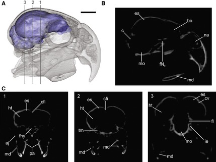
Figure 1.

Three‐dimensional reconstruction of Cyanoliseus patagonus endocast, generated from CT scans. (A) Lateral view of the skull in transparence showing the endocast; (B, C) tomographs of the skull in (B) sagittal and (C) coronal planes (1–3) showing the spaces occupied by the brain and its different regions/structures. aj, arcus jugalis; bo, bulbus olfactorius; c, cerebellum; cfi, crista frontalis interna; cv, crista vallecularis; es, eminentia sagittalis; fhy, fossa hypohysialis; fl, floculus; fNII, foramen nervus opticum; ht, hemisphaeria telencephali; ie, inner ear; md, mandible; mo, medulla oblongata; na, apertura nasi ossea; pa, ossa palatina; tm, tectum mesencephali. Scale bar: 1 cm.
