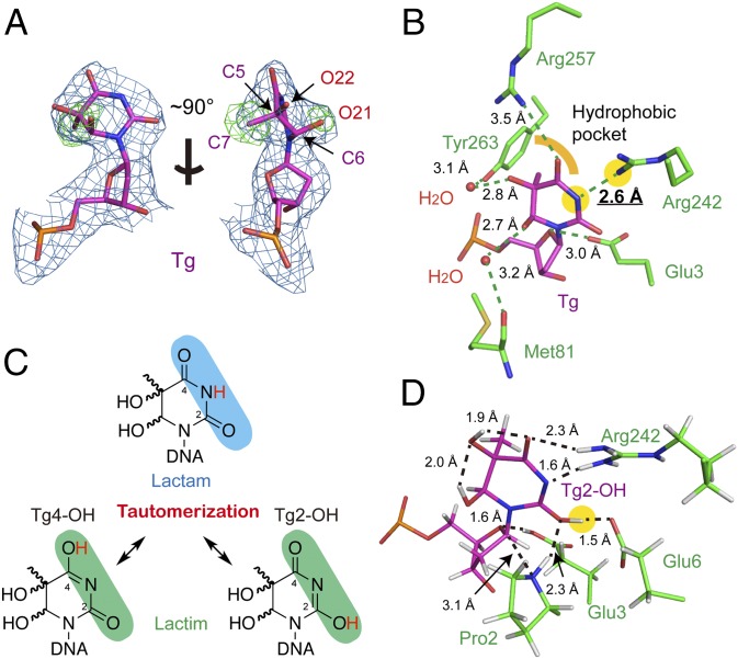
Fig. 2.
Tautomerization-dependent recognition of Tg. (A) Electron density map of Tg. The blue 2Fobs − Fcal map is contoured at 1.2σ and the green Fobs − Fcal omit map—by removing the 5-methyl (C7) and 6-hydroxyl group (O21) of Tg—is contoured at 3.0σ [omit maps removing 5-hydroxyl group (O22) are shown in SI Appendix, Fig. S3A]. (B) The active-site pocket of Tg-bound NEIL1. Hydrogen bonds are shown in green dashed lines. The hydrophobic pocket surrounding the 5-methyl group of Tg is indicated by a yellow curve. The N3 of Tg and Nη of Arg242 is highlighted with yellow background. (C) Tg tautomers in the lactam (Upper) and lactim (Lower) forms. (D) Optimized structure of the Tg-bound NEIL1 active site. Due to the Tg2-OH tautomer, a new hydrogen bond was observed between 2-OH of Tg and Glu6. Key distances are marked in black (in angstroms), and the 2-OH group of Tg is highlighted with yellow background.

