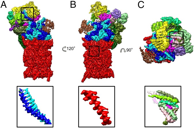
Fig. 1.
Cryo-EM reconstruction of the human 26S proteasome at 3.9 Å resolution. (A–C) Three different views of the 26S proteasome colored according to its subunits. Red: CP; blue: AAA-ATPase heterohexamer; brown: Rpn1; yellow: Rpn2; green: PCI subunits (Rpn3, -5, -6, -7, -9, -12); magenta: Rpn8, -11; purple: Rpn10. (Bottom) Selected, magnified features (coiled-coil Rpt3/6, helix formed by residues 57–80 of β1, a helical bundle from the lid subcomplex) from the marked areas.

