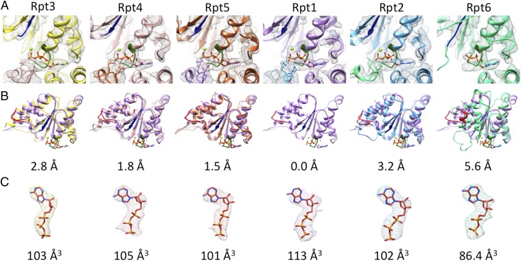
Fig. 3.
Nucleotide binding and structures of large AAA subdomains. (A) Nucleotide densities and coordinated Mg2+ and Arg–fingers of the neighboring subunits at the Walker A motifs (green) of the Rpts. (B) Structural comparison of the large AAA+ domain of each Rpt with Rpt1 (Walker A green, Walker B dark blue, pore loop red). Below each panel the root mean squared deviation of the respective structure compared with Rpt1 in angstroms is assigned. (C) EM densities of bound nucleotides and modeled nucleotides. Below each panel the volume of the difference map in Å3 (cubic Angstroms) is shown.

