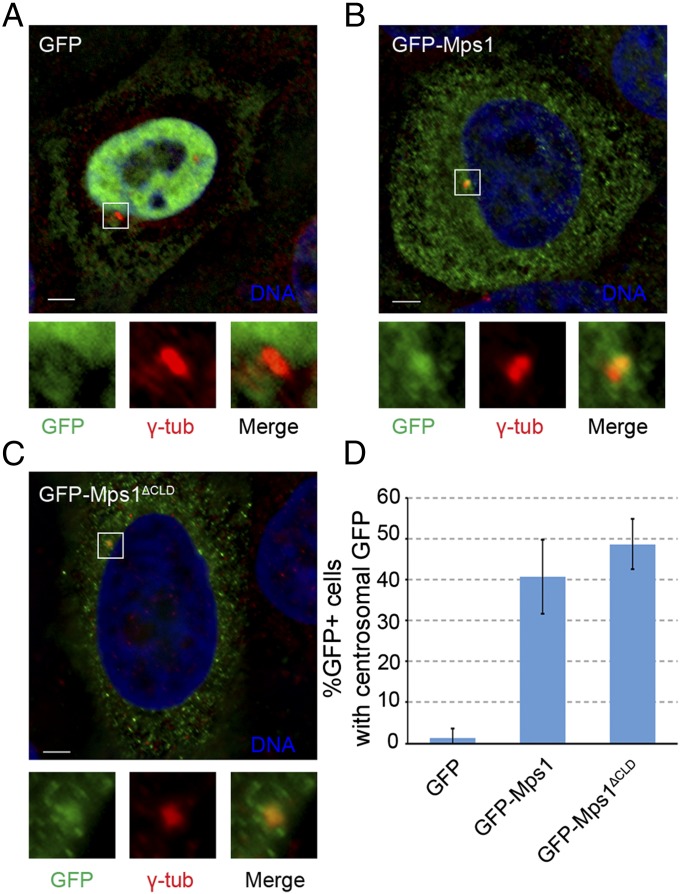
Fig. 1.
The CLD is not necessary for Mps1 centrosomal localization. (A–C) Representative images of S-phase arrested HeLa cells transfected with GFP (A), GFP-Mps1 (B), or GFP-Mps1ΔCLD (C), showing DNA (blue), GFP (green), and centrosomes (γ-tub, red). In this and subsequent figures, lower panels depict approximate fourfold digital magnification of boxed regions. (Scale bars, 5 µm.) (D) Percentage of GFP+ cells in which GFP colocalizes with γ-tubulin; mean ± SD (SD) of n = 3 independent experiments of n = 50 cells for each experiment.

