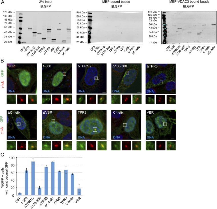
Fig. S1.
TPR3 and downstream elements are required for VDAC3 binding and centrosomal localization. (A) In vitro pulldown assay between indicated constructs expressed in HEK293 cells and either MBP (Center) or MBP-VDAC3 (Right). MBP or MBP-VDAC3 were expressed in Escherichia coli and bound to amylose resin, and the ability of each construct to bind to the amylose resin was assessed by immunoblotting with an antibody against GFP. (Left) Two percent of the amount of lysate added to the amylose resin. Size of molecular markers for each blot is indicated in kilodaltons. (B) Representative images of asynchronous HeLa cells transfected with the indicated constructs, showing DNA (blue), GFP (green), and centrosomes (γ-tub, red). In this and subsequent figures, lower panels depict approximate fourfold digital magnification of boxed regions. (Scale bar, 5 µm.) (C) Quantification of the percentage of GFP+ cells where GFP signal colocalizes with γ-tubulin at centrosomes, presented as the mean ± SD (SD) of n = 3 independent experiments of n = 50 cells for each experiment; error bars indicate SD (SD).

