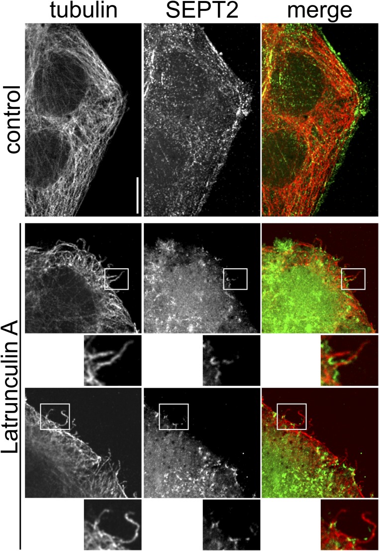
Fig. S6.
Formation of microtubule-based protrusions with septin base after latrunculin A treatment. Indirect immunofluorescence of SEPT2 (green) and α-tubulin (red) in Caco-2 cells. Cells were treated with 5 nM latrunculin A for 60 min. Treatment with latrunculin A causes similar septin accumulations at the base of protrusions as a treatment with CDT does. Magnifications of Insets are below the images. (Scale bar, 5 µm.)

