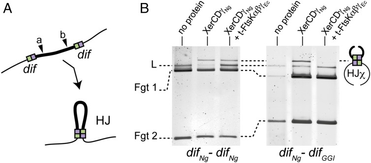
Fig. 5.
In vitro HJ formation. (A) Schematic of the in vitro HJ formation assay using a color code as in Fig. 1. NcoI restriction site is represented: (a) for difNg-difNg cassette and (b) for difNg-difGGI cassette. (B) HJ formation for difNg-difNg (Left) or difNg-difGGI (Right) cassettes. As indicated, DNA substrates were incubated with XerDNg and XerDγNg ± t-FtsKαβγEc. After DNA restriction, HJχ were separated from substrate DNA by electrophoresis: linear (L, partial restriction) and fragments (Fgt 1 and Fgt 2, total restriction).

