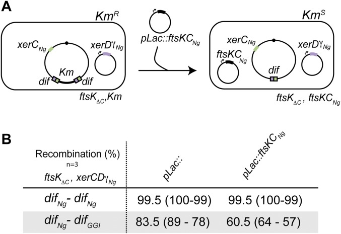
Fig. S3.
In vivo recombination reaction. (A) Schematic of the assay. The color code is as in Fig. 1. (B) In vivo recombination reactions using E. coli strains containing the difNg-difNg or difNg-difGGI cassette inserted at the dif locus. Recombination was scored as the percentage of kanamycin-sensitive colonies: mean (minimum – maximum) (Methods).

