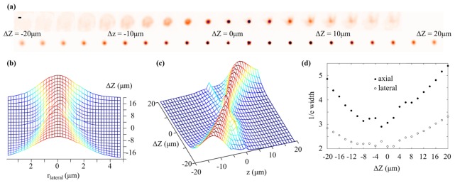
Fig. 3.
(a) Images of a 2.5 μm diameter green fluorescent microsphere acquired as the objective lens is displaced axially. Top row: conventional widefield images. Bottom row: in-focus plane extracted from reconstructed light field focal series. Scale bar in upper left corner is 3 µm. (b) Lateral and (c) axial line profiles taken through the centre of the bead in the focal series reconstruction as the objective lens is axially displaced. (d) Lateral (unfilled circles) and axial (filled circles) 1/e width of the bead image in the focal series reconstruction as the objective lens is displaced axially.

