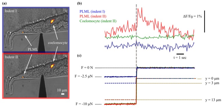
Fig. 6.
(a) DIC images showing indentation of a C.elegans specimen to depth of approximately 3 μm (top) and 13 μm (bottom) with a microforce sensing probe approximately 110 μm from the tip of the tail. Maximum intensity projection of reconstructed light field (displayed with a red hot colour map) overlaid to show positions of PLML and coelomocyte. (b) Total brightness of the cell body of the PLML indicates that the TRN is only activated by the withdrawl of the probe following the larger indentation (red line). The signal from a nearby GFP transgenic marker (green line) is constant despite similar displacement following withdrawal of the probe. (c) Corresponding force (solid lines) and probe position (dashed lines) measured during the end of the hold phase of indentation and after probe retraction.

