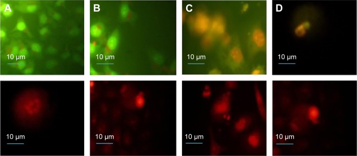
Figure 4.
Fluorescence microcopy images of HeLa cells stained with AO (first row) and EB (second row) after exposure to NOP-DOX@BSA-FA and a light dose.
Notes: (A) Cells exposed for 5 minutes showing the formation of apoptotic bodies within the cells; (B) apoptotic and necrotic cells at 15 minutes, with 50% of the cells showing necrosis and the remaining showing early and late apoptosis; and (C, D) apoptotic and necrotic cells at 30 minutes exposure showing nuclear condensation, budding to form apoptotic bodies, and nuclear fragmentation. Magnification is 60×.
Abbreviations: AO, acridine orange; BSA, bovine serum albumin; DOX, doxorubicin; EB, ethidium bromide; FA, folic acid; HeLa, human cervical epithelial malignant carcinoma; NOP, nickel oxide nanoparticle.

