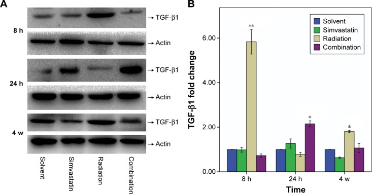
Figure 5.
Protein content of TGF-β1 (25 Kd) in SMGs of mice at 8 hours, 24 hours, and 4 weeks after IR was assessed by Western blot analysis.
Notes: (A) Representative Western blots from three to five experiments with similar results. (B) Changes in TGF-β1 quantified by scanning densitometry analysis using Image Lab software. The data (relative density normalized to β-actin) are expressed as mean ± SD; *P<0.05, **P<0.01.
Abbreviations: TGF-β1, transforming growth factor β1; SMG, submandibular gland; IR, irradiation; SD, standard deviation; h, hours; w, weeks.

