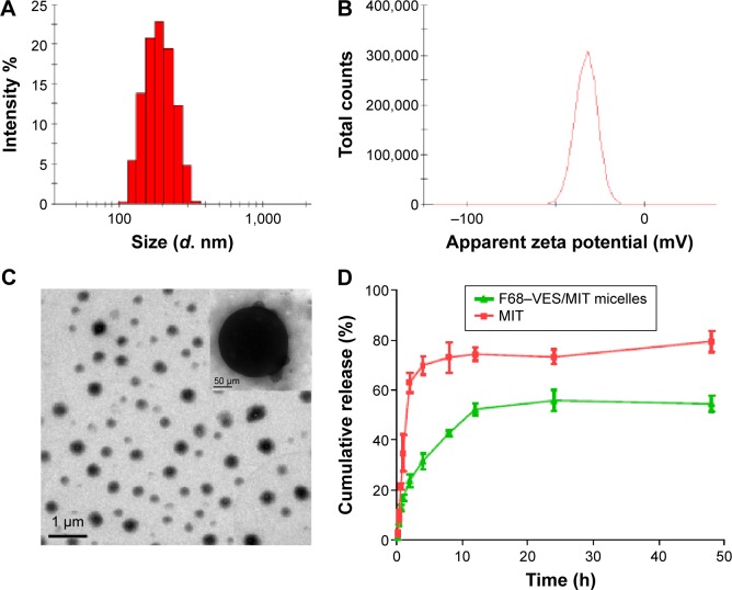
Figure 4.
Characterization of F68–VES/MIT micelles.
Notes: (A) Size distribution of F68–VES/MIT micelles. (B) Zeta potential of F68–VES/MIT micelles. (C) TEM images of F68–VES/MIT micelles. (D) In vitro accumulative release of MIT from free MIT solution and F68–VES/MIT micelles. Error bar represents the standard deviation value of three experiments.
Abbreviations: F68–VES/MIT micelles, mitoxantrone-loaded Pluronic F68-conjugated vitamin E succinate polymer micelles; MIT, mitoxantrone; TEM, transmission electron microscopy.

