
Fig.2. Vessel malformations and tumor growth are enhanced in TRPV4 knockout mice.
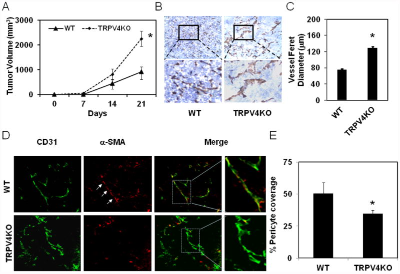
A) Time-dependent growth of the tumors in WT and TRPV4KO mice. Mouse Lewis lung carcinoma (LLC) cells (2 × 10⁶) were subcutaneously injected in to wild type C57BL/6 mice (WT) or TRPV4 knockout mice in C57BL/6 background (TRPV4KO) and tumor growth was measured using calipers at indicated days. The data shown are ± SEM of three independent experiments (n=8-10 mice for each group). B) Immunohistochemical analysis showing increased vessel diameter (feret) in tumors (21 days) from TRPV4 knockout mice (TRPV4KO) compared to wild type mice (WT). C) Quantitative analysis of microvessel diameter in tumors from WT and TRPV4KO mice. D) Frozen sections of tumors (10 μm thickness) were stained with CD31 (green) and α-SMA (red) to measure pericyte coverage (matured vessels). E) Quantitative analysis of pericyte covered microvessels in tumors from WT and TRPV4KO mice. The results shown are mean ± SEM from 3 independent experiments. The significance was set at p≦ 0.05.