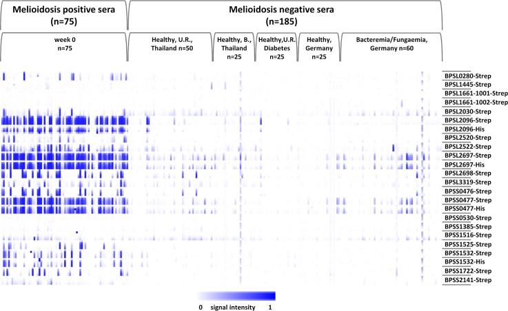
Fig 2. Heatmap of probing a collection of melioidosis-positive sera and negative control sera.
Protein arrays containing 20 B. pseudomallei recombinant proteins were probed with 260 melioidosis and nonmelioidosis sera. The melioidosis positive sera (n = 75) were drawn at week 0 (p.a.) from patients with B. pseudomallei infections. All positive sera were sampled in Ubon Ratchathani, Thailand. Negative control sera of healthy persons (n = 125) were sampled in the endemic regions of Thailand ((Ubon Ratchathani (U.R.) and Bangkok (B.)), Thailand and non-endemic region of Greifswald, Germany. Additionally, further negative control sera of patients with other bacteremia or fungaemia (n = 60) were used from the non-endemic region of Greifswald. Not shown are the results of incubations with meliodosis-positive sera obtained 12 and 52 weeks after admission. The antigens are shown in rows with five increasing concentrations per protein, and the patient samples are represented in columns. Array signals are reflected by the intensities of the color (white to blue) inside the boxes. The heatmap was created using Multi experiment Viewer (MeV 4.9.0) from TM4 suite, USA.

