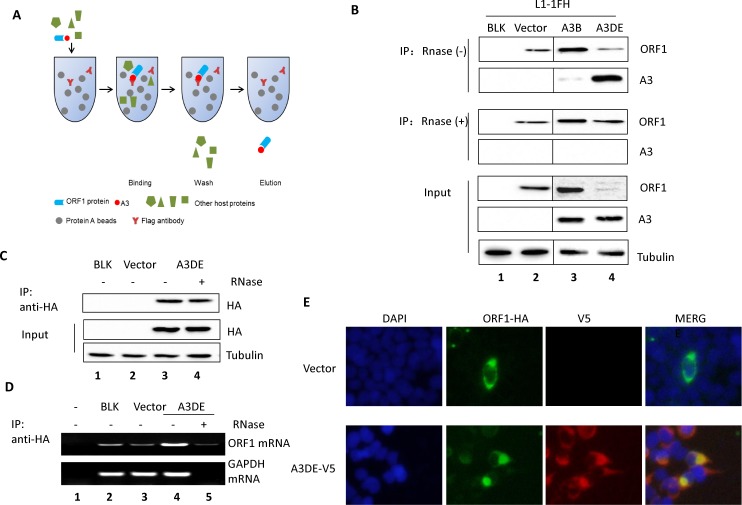
Fig 3. APOBEC3DE interacts with ORF1p in a RNA-dependent manner.
(A) Diagram of the co-IP assay. Cleared whole-cell lysate was used for these assays. Protein antigens were pulled down by specific antibodies. The antibodies were coupled to solid substrates. (B) A3B or A3DE interacts with ORF1p, and RNase disrupts the binding of A3F to ORF1p. The pc-L1-1FH plasmid was co-transfected with A3B-V5 or A3DE-V5 or the control vector into HEK293T cells. The cleared cell lysate was split into two halves, and RNase A was added to one half (final concentration, 50 μg/ml); both samples were then incubated with Flag-tagged beads. Western blotting was performed to identify the input, RNase A-treated, and untreated IP products. (C, D) IP of A3DE for endogenous L1 RNP. A3DE-HA was transfected into HEK293T cells and HA beads were used to pull down A3DE. IP assay was conducted at 48 hours post-transfection. IP product was aliquoted, one for Western Blotting and the other one for RNA extraction and L1 mRNA detection. (E) Immunofluorescence staining of A3DE and ORF1p in HEK293T cells.

