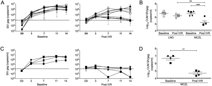
Fig 3. Tissue-associated MIV-150 inhibits ex vivo SHIV-RT infection of vaginal and cervical mucosa.
MZCL (n = 5) or LNG (n = 4) IVRs were inserted for 24h. Vaginal and ectocervical biopsies were collected immediately after IVR removal (Post) and at the baseline. Non-stimulated vaginal (n = 2–6) and cervical (n = 2–4) explants were challenged with SHIV-RT (104 TCID50/explant) for ~18h, washed and cultured for 14d with IL2. Infection was monitored and analyzed as in Fig 1. Shown are (A) SIV gag copies/ml of each animal (Mean of replicates ± SEM; symbols match those shown in panel B) and (B) SIV gag CUM analyses of Log10 transformed data (Mean ± SEM) in vaginal tissues. Each symbol represents an individual animal (MZCL group: closed symbols; LNG group: open symbols). Shown are (C) SIV gag copies/ml of each animal (Mean of replicates ± SEM; symbols match those shown in panel D) and (D) SIV gag CUM analyses of Log10 transformed data (Mean ± SEM) in cervical tissues. Each symbol represents an individual animal (MZCL group). Significant p-values of <0.0001(***) and <0.001(**) are indicated. Input SIV gag copy numbers (Mean; post washout after challenge) are shown as dotted lines.

