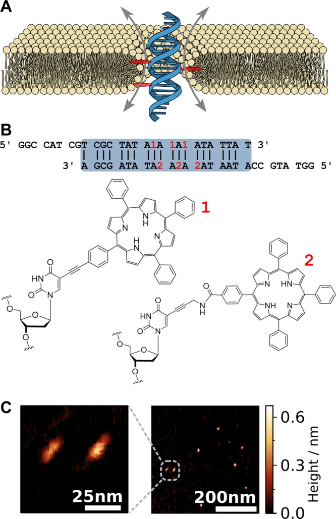
Figure 1.

Design, shape, and dimensions of the membrane-spanning DNA duplex. (A) Envisioned placement and conductance mechanism of the DNA duplex (blue) decorated with six porphyrin membrane anchors (red, only three shown in cross section) causing the formation of a toroidal lipid pore (yellow). The sketch is roughly drawn to scale. (B) The nucleotide sequence of the duplex. The locations of the porphyrin tags within the sequence are indicated in red. The bottom row shows the chemical structures of the tags labeled as 1 (with acetylene linker) and 2 (with amide linker) according to the target strand. (C) AFM images of the duplex structures adsorbed onto mica and imaged in air.
