
Figure 5. In vivo.
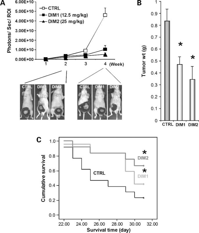
anticarcinogenic activity of DIM-C-pPhCl. A, effects of DIM-C-pPhCl on primary bladder tumors. Three different treatment groups consist of DMSO control (CTRL; n = 14), 12.5 mg/kg/d DIM-C-pPhCl (DIM1; n = 13), and 25 mg/kg/d DIM-C-pPhCl (DIM2; n = 13). Each group was treated thrice a week right after randomization. 253J B-V cells (2 × 10⁵) were implanted into bladder wall of male nude mice. Mice were injected with luciferin i.p. and were imaged on the charge-coupled device camera 10 min after injection. Bioluminescent imaging images were collected for 1 or 10 s for each group. Different image acquisition times were needed to avoid saturating the charge-coupled device camera. Bioluminescence is presented as a pseudocolor scale: red, highest photon flux; blue, lowest photon flux. Bioluminescence from primary tumors was quantified by region of interest analysis of images obtained on the indicated time points (X axis) after cell implantation. Background bioluminescence was subtracted from each tumor region of interest value. Mean ± SE photon flux in each group. Bottom, representative bioluminescent imaging images of primary 253J B-V bladder tumors 2 and 4 wk after initial treatment. B, tumor size measurements. Actual tumor sizes were measured 4 wk after implantation of cancer cells. Mean ± SE tumor volume. *, P < 0.05. C, effects of DIM-C-pPhCl on human bladder xenograft mice survival. Kaplan-Maier plots were generated, and survival time of animals was analyzed for significance by log-rank survival analysis. *, P < 0.05.