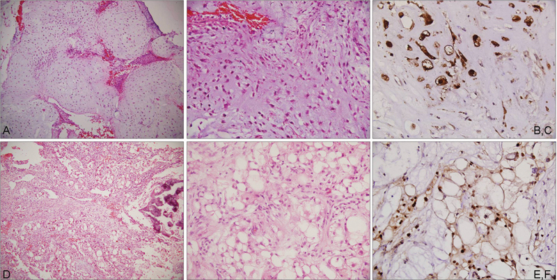
Fig. 1.

Photomicrographs showing a chondrosarcoma composed of cartilaginous lobules (a; HE, ×100) with high cellularity and pleomorphism (b; HE, ×200), immunopositive for S-100 (c; IHC, ×400); chordoma with a myxoid matrix (d; HE, ×100) small polygonal cells and large physaliphorous cells (e; HE, ×200) immunopositive for brachyury (f; IHC, ×400). HE, hematoxylin and eosin stain; IHC, immunohistochemistry.
