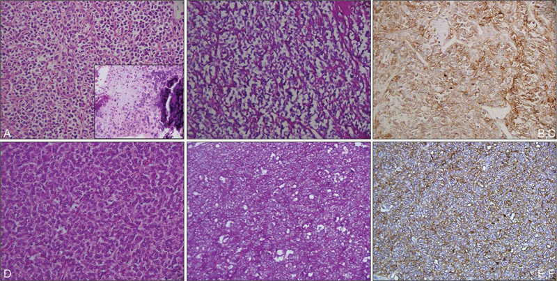
Fig. 2.

Photomicrographs showing mesenchymal chondrosarcoma with predominant malignant small round cell areas with occasional foci of well differentiated cartilage (inset) (a; HE, ×200); small round cells are negative on PAS stain (b; PAS, ×200) and are immunopositive for CD99 (c; IHC, ×200); case of Ewing sarcoma showing a malignant small round cell tumor (d; HE, ×200); with cytoplasmic PAS-positive glycogen (c; PAS, ×200) and CD99 immunopositivity (f; IHC, ×200). HE, hematoxylin and eosin stain; IHC, immunohistochemistry; PAS, periodic acid-Schiff.
