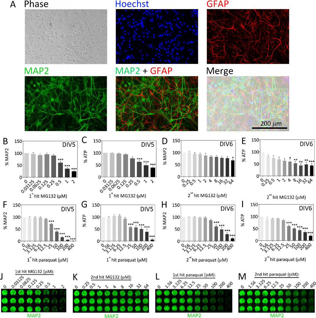
Figure 1. Characterization of primary hippocampal cultures and concentration-response curves.
(A) Hippocampal cultures harvested from the postnatal rat brain were immunocytochemically stained on day in vitro 7 (DIV7) for the neuronal marker microtubule-associated protein 2 (MAP2, green) and the astrocyte marker glial fibrillary acidic protein (GFAP, red). Nuclei were stained blue with the Hoechst reagent. Photomicrographs were captured using a 20× objective on an epifluorescent microscope. (B–M) Primary hippocampal cultures were treated with MG132 or paraquat on DIV5 (1st hit) or DIV6 (2nd hit). Viability was assayed on DIV7 by (B, D, F, H) the In-Cell Western assay for MAP2 levels and (C, E, G, I) the Cell Titer Glo luminescent assay for ATP. Representative MAP2 In-Cell Western images are shown in panels J–M. Shown are the mean and SEM of 3–4 independent experiments, each performed in triplicate. *p ≤ 0.05, **p ≤ 0.01, ***p ≤ 0.001 versus 0 μM MG132 or paraquat, one-way ANOVA followed by Bonferroni post hoc correction.

