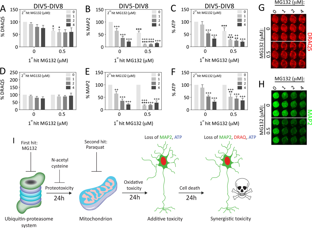
Figure 6. Impact of dual hits of MG132 on hippocampal neuron viability.
Primary hippocampal cultures were treated on DIV5 with the 1st hit of MG132 and on DIV6 with a range of concentrations of MG132 as the 2nd hit. Viability was assayed 48h later on DIV8 by (A) the nuclear stain DRAQ5, (B) the In-Cell Western assay for MAP2 levels, and (C) the Cell Titer Glo luminescent assay for ATP. Representative DRAQ5 and MAP2 images are shown in panels G and H. Data are expressed as a function of the 0 μM 2nd hit group in panels D–F to statistically evaluate the effect of dual hits. The loss of viability in response to dual hits of MG132 was synergistic, but only at one concentration of the 2nd hit (1 μM) and not according to the DRAQ5 nuclear assay. Shown are the mean and SEM of 3–5 independent experiments, each performed in triplicate. *p ≤ 0.05, **p ≤ 0.01, ***p ≤ 0.001 versus 0 μM 1st hit; +p ≤ 0.05, ++p ≤ 0.01, +++p ≤ 0.001 versus 0 μM 2nd hit; two-way ANOVA followed by Bonferroni post hoc correction. (I) Summary schematic for the major findings in the present study. The images of the proteasome and mitochondrion are adapted from our previous drawings (Anne Stetler et al., 2013; Leak, 2014).

